Technology
From volume to validated report.
SPView combines 3D segmentation, reproducible quantification and structured report drafting. Automation supports the workflow; the radiologist retains responsibility for interpretation and validation.
Principles
What happens between the scan and the report.
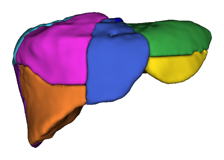
Segmentation
AI-assisted identification of anatomical structures in CT and MRI imaging.
Quantification
Volumes, diameters, locations and anatomical relationships are extracted from the segmented 3D volume.
Structured reporting
Quantitative findings become an editable draft report. The radiologist reviews and validates the final report.
Technology
Advanced AI.
Designed for clinical reality.
SPView segments CT and MRI volumes in 3D, extracts measurements and drafts structured findings. The radiologist reviews the anatomy, interprets the findings and validates the final report.
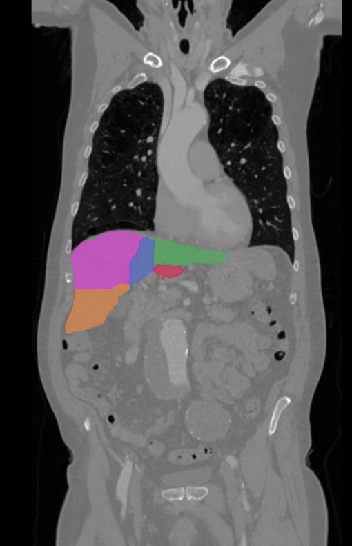
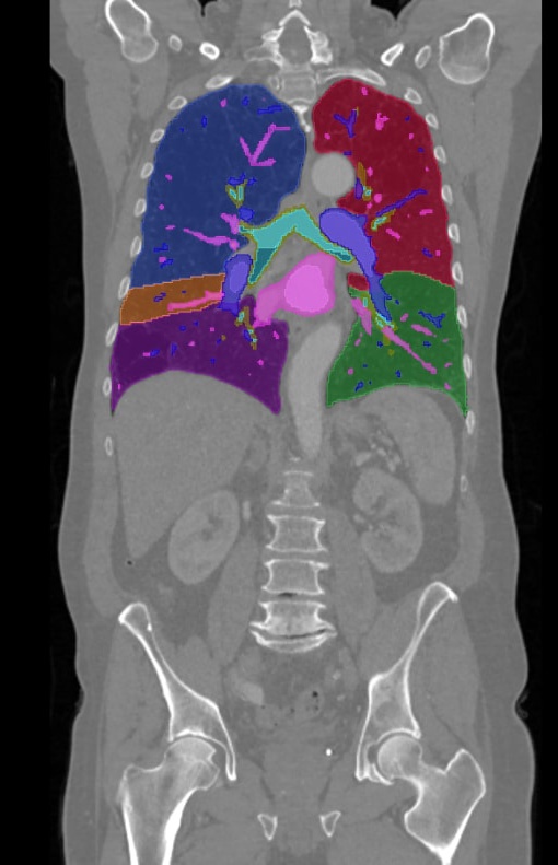
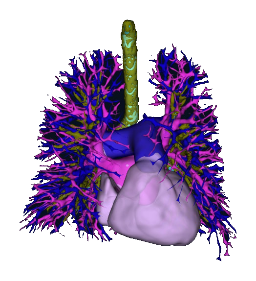
See your next scan become a report.
Explore 3D reconstruction, quantitative measurements and structured draft reports — with radiologist review and validation at the center.