Lung module
AI-powered 3D planning for thoracic surgery.
SPview reconstructs bronchovascular anatomy from the patient's CT examination, supporting patient-specific visualization of the structures relevant to thoracic surgical planning.
Runs locally on the SPview workstation.
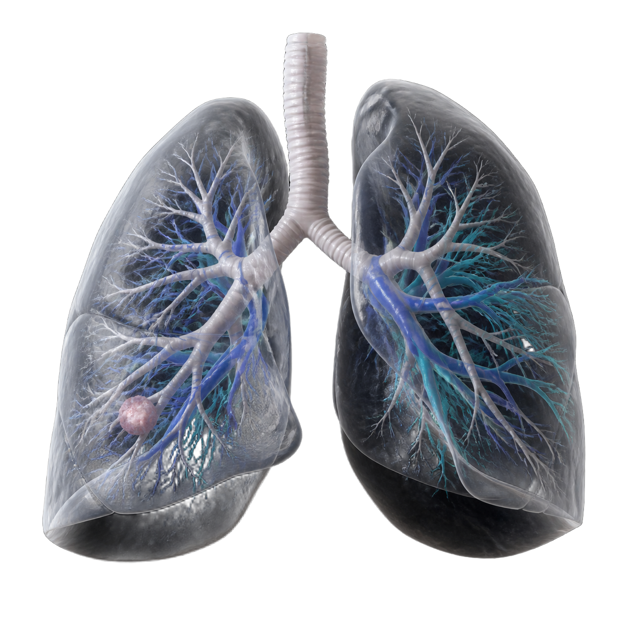
Reconstructed structures
Bronchovascular anatomy in three dimensions.
Lung anatomy
Lungs and lobar organization reconstructed from the patient's CT.
Bronchi
Bronchial tree visualized in relation to the parenchyma.
Pulmonary arteries
Arterial branching relevant to anatomical resection.
Pulmonary veins
Venous return anatomy in three dimensions.
Lesions
Lesion position relative to bronchovascular structures.
Anatomical relationships
Spatial context for the planned surgical approach.
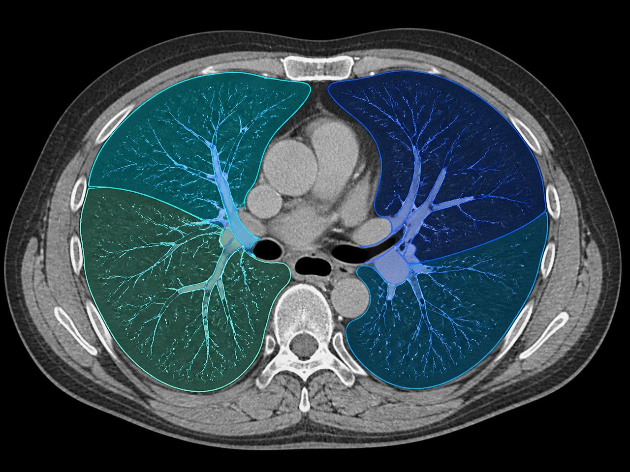
From CT to 3D
Imaging in. Anatomy out.
Thoracic examinations are processed on the SPview workstation inside the hospital, without transferring patient DICOM files to an external platform.
- 01Chest CT examination
- 02SPview workstation
- 03AI-assisted lung segmentation
- 04Interactive 3D lung model
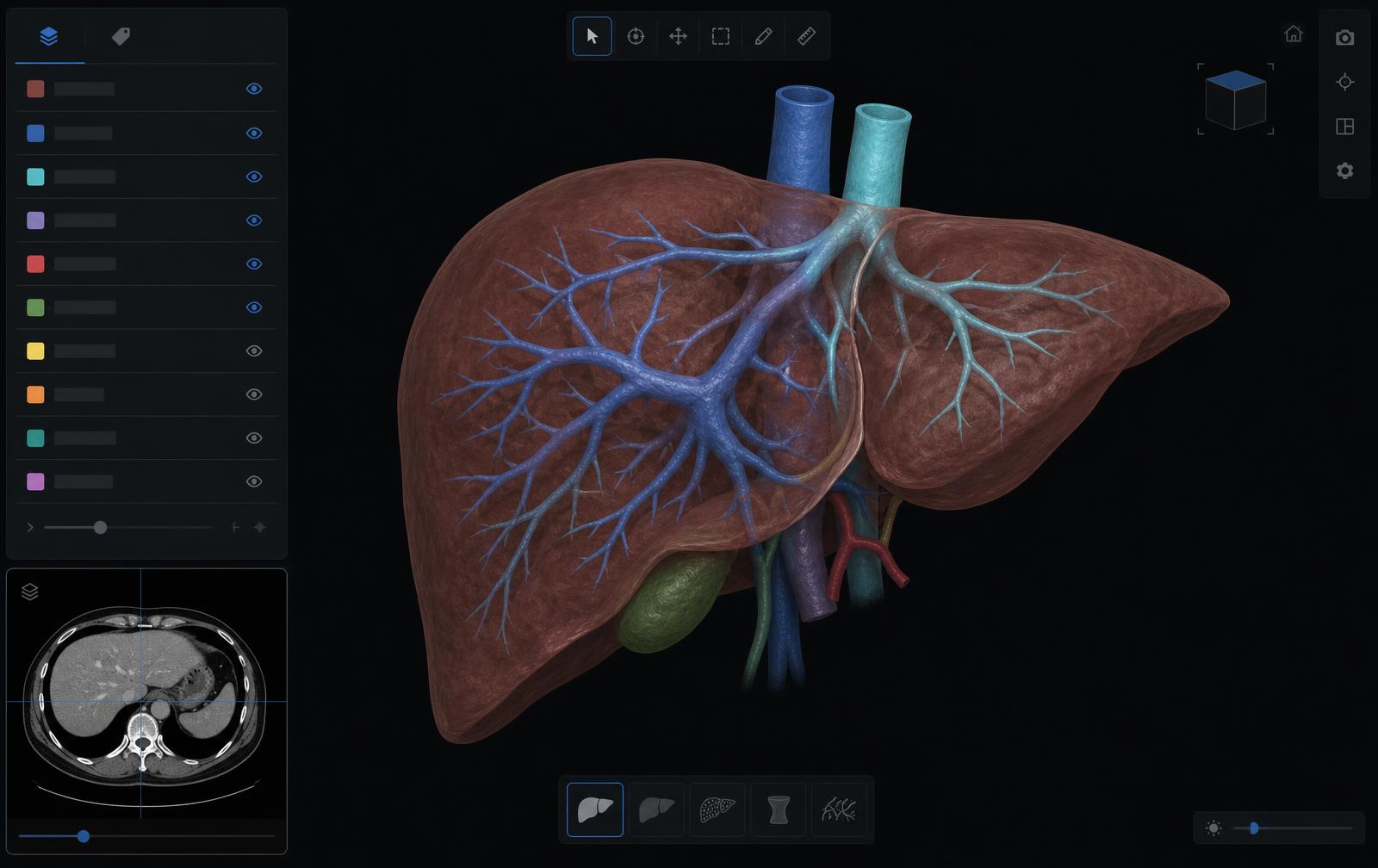
Software
Vessels and bronchi, on demand.
Toggle structures individually to study lesion position and bronchovascular relationships.
See SPview Lung
on your own cases.
Request a demonstration of the Lung module on the SPview workstation, inside your institution.
Dedicated workstation. Local processing. Liver + Lung.