Liver module
AI-powered 3D planning for liver surgery.
SPview reconstructs patient-specific hepatic anatomy from CT or MRI imaging, so the surgical team can explore parenchyma, vasculature and lesions in three dimensions before entering the operating room.
Runs locally on the SPview workstation.
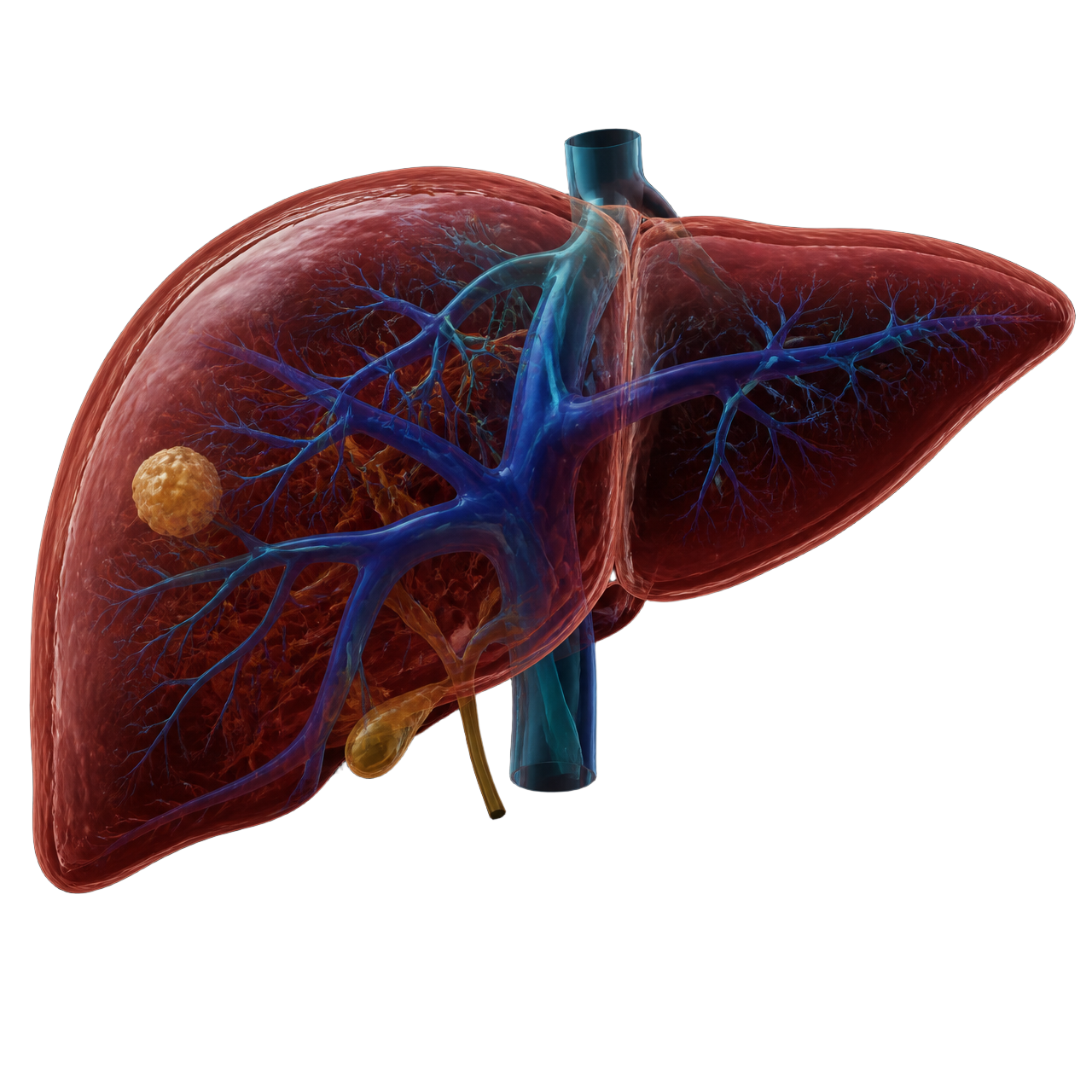
Reconstructed structures
Hepatic anatomy, structure by structure.
Liver parenchyma
Volume and surface of the liver reconstructed from the patient's imaging.
Hepatic veins
Drainage anatomy visualized in relation to the parenchyma.
Portal venous anatomy
Portal branching relevant to anatomical resection planning.
Lesions
Position of lesions relative to vascular structures.
Anatomical segmentation
Segmental organization of hepatic anatomy.
From CT to 3D
Imaging in. Anatomy out.
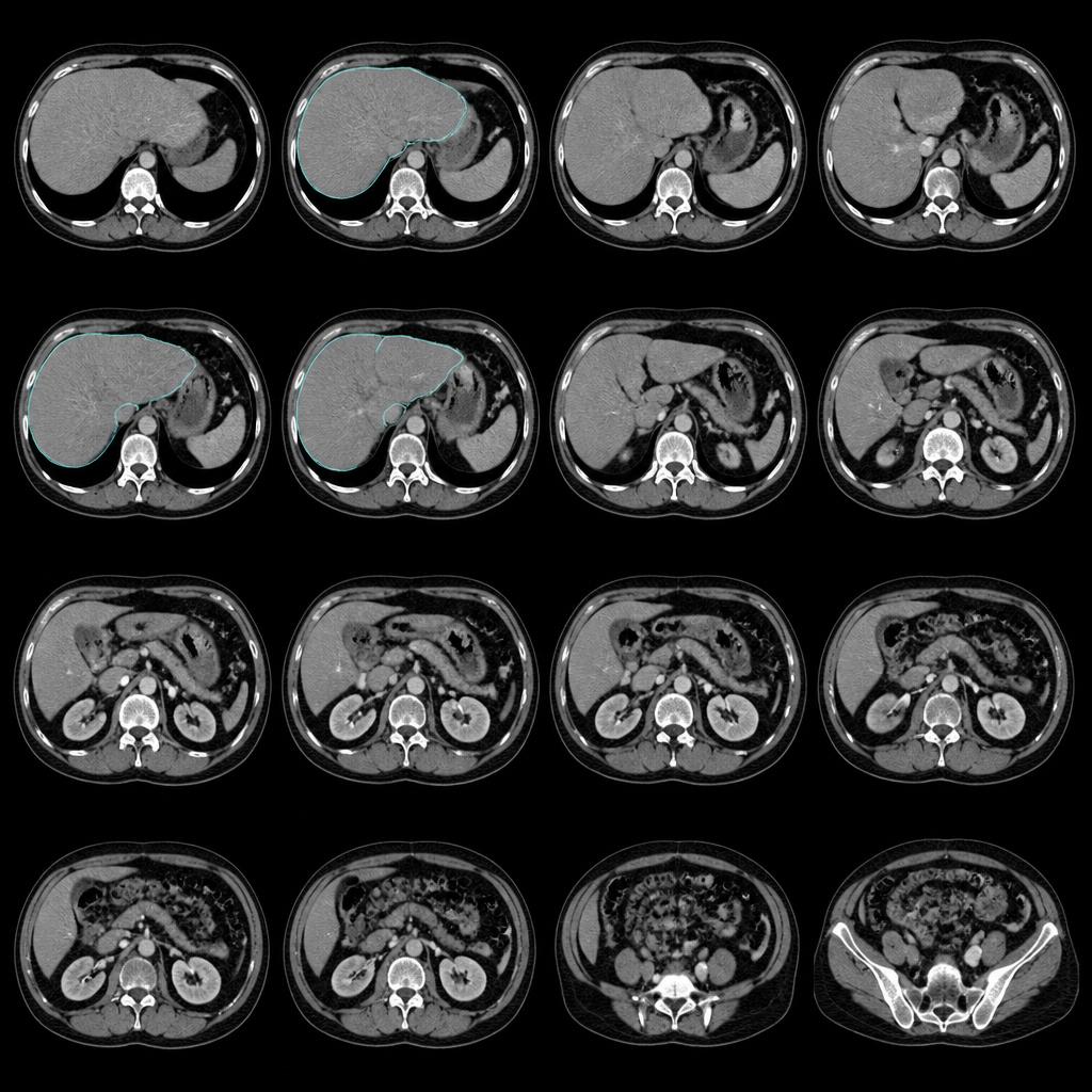
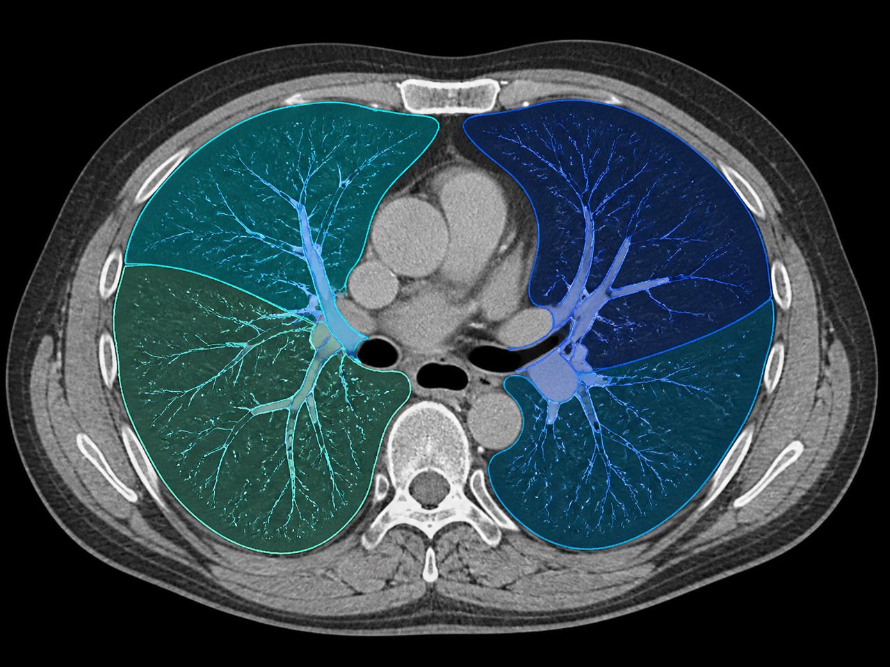
The examination is processed on the SPview workstation inside the hospital. No DICOM export to an external platform, no anonymization workflow before processing.
- 01CT / MRI examination
- 02SPview workstation
- 03AI-assisted liver segmentation
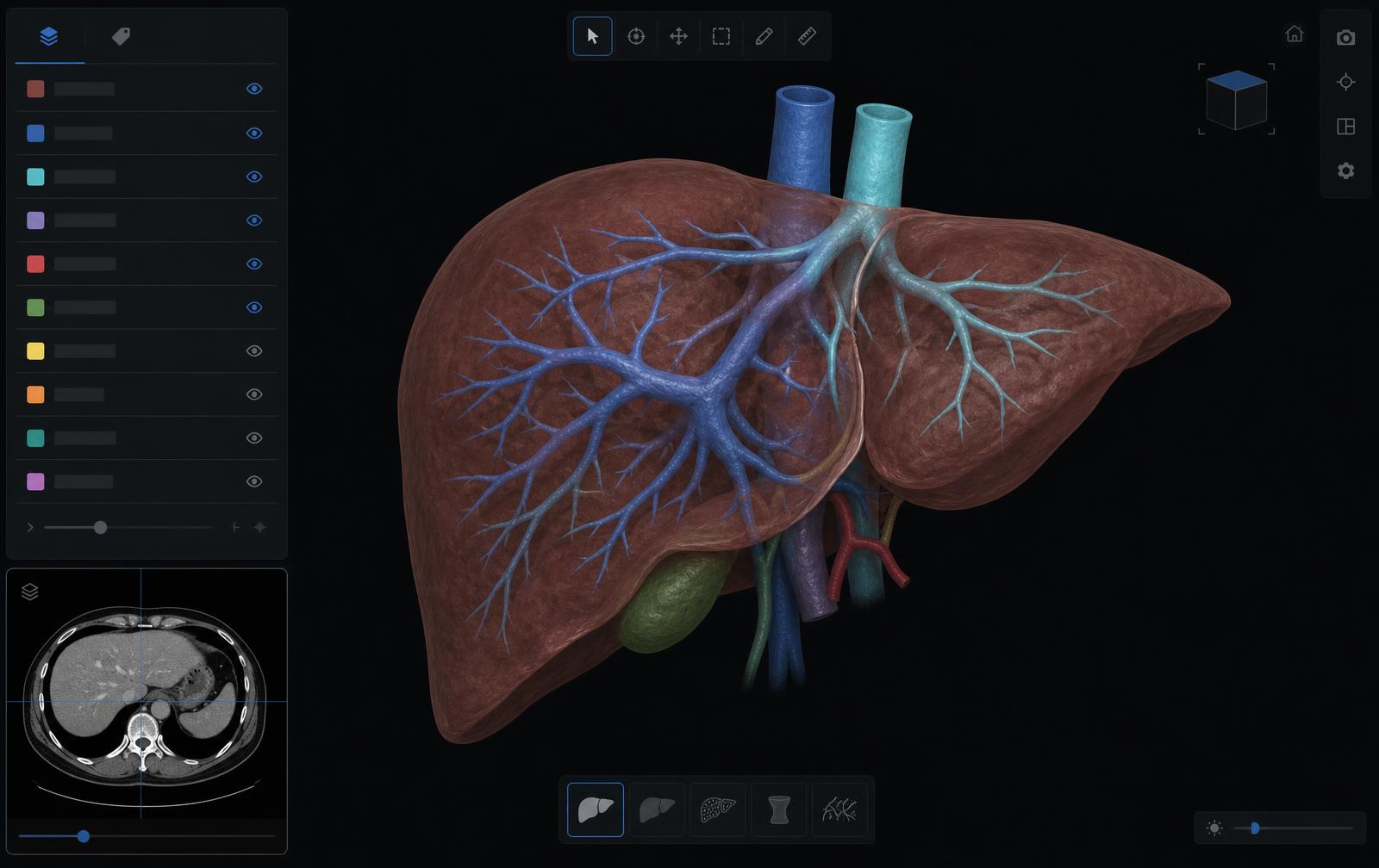
- 04Interactive 3D liver model
Software
Explore the reconstruction interactively.
Rotate, zoom and toggle anatomical structures to study the relationships that matter for the planned approach.
See SPview Liver
on your own cases.
Request a demonstration of the Liver module on the SPview workstation, inside your institution.
Dedicated workstation. Local processing. Liver + Lung.