AI-powered surgical planning
See the anatomy.
Plan with confidence.
SPview uses artificial intelligence to transform medical imaging into patient-specific 3D anatomical models, helping surgeons explore complex anatomy before surgery.
Dedicated hardwareLocal processingNo external DICOM transfer
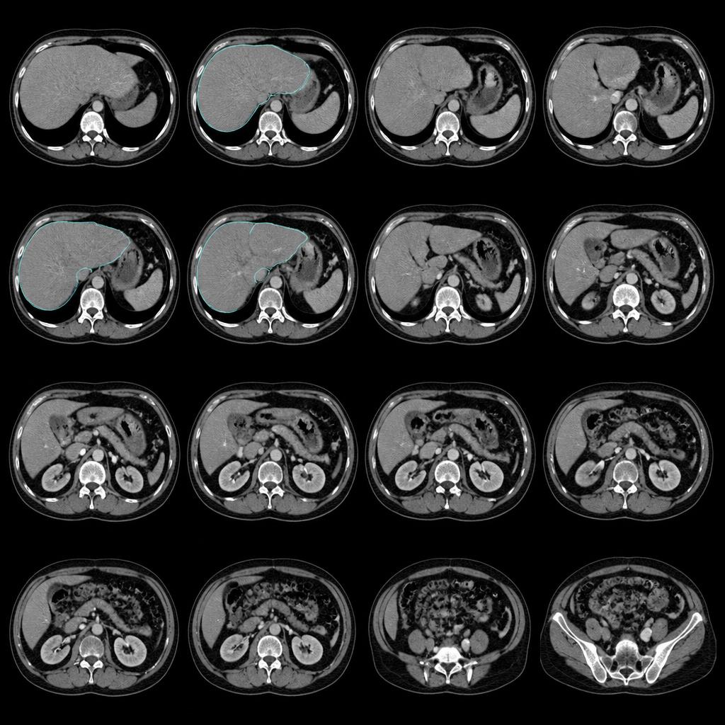
Imaging to anatomy
2D imaging.
3D anatomy.
Surgeons often need to mentally reconstruct complex three-dimensional anatomy from two-dimensional CT or MRI images. SPview transforms this imaging information into an intuitive patient-specific 3D representation.
One software environment
One platform.
Two surgical specialties.
Liver and Lung modules are available within the same SPview software environment, allowing different surgical departments to access advanced 3D planning from one solution.
One workstation. One software. Liver + Lung.
How it works
From scan to surgical insight.
Medical imaging
Access the patient's CT or MRI examination directly within the hospital workflow.
DICOM / CT / MRI
AI-assisted segmentation
SPview identifies and reconstructs relevant anatomical structures.
AI processing
Interactive 3D model
Explore patient-specific anatomy in three dimensions and use it to prepare the surgical strategy.
3D model
Local workflow
No DICOM transfers.
No anonymization workflow.
No unnecessary complexity.
SPview provides the dedicated workstation and the software already installed. Medical imaging can be processed within the hospital environment without sending patient DICOM files to an external platform and without the usual time-consuming anonymization and transfer process.
Traditional external workflow
- 01CT / MRI
- 02Export DICOM
- 03Anonymize patient data
- 04Upload / transfer files
- 05External processing
- 06Retrieve the result
- 07Review the reconstruction
Seven steps before the surgical team can look at the anatomy.
SPview workflow
- 01CT / MRI
- 02SPview workstation
- 03AI reconstruction
- 04Interactive 3D model
From medical imaging to 3D surgical planning without sending patient data outside your institution.
Your imaging stays where it belongs: inside your hospital.
Hardware + software
Everything you need.
Ready to use.
SPview can be delivered as a complete clinical solution combining dedicated high-performance hardware and the SPview software. The workstation is configured for AI-assisted 3D reconstruction and arrives with the SPview environment installed.

SPview workstation
SPview software
Ready-to-use 3D surgical planning
Ready to deploy
Dedicated hardware configured for SPview.
Local clinical workflow
Patient imaging does not need to be transferred to SPview for external processing.
Liver + Lung
Both clinical modules can run within the same SPview software and workstation, enabling different surgical departments to use the same solution.
Technology
Advanced AI.
Designed for clinical reality.
Complex segmentation technology should not create additional complexity for clinicians. SPview combines AI-assisted medical image segmentation with a workflow designed around surgeons, radiologists and hospital environments.
Built for surgeons
Understand complex anatomy before the first incision.
Patient-specific anatomy
Visualize each patient's unique anatomical relationships.
Spatial understanding
Explore vessels, lesions and surrounding structures in three dimensions.
Pre-operative planning
Use the reconstruction to prepare and discuss the surgical approach.
Communication
Use intuitive 3D visualization when discussing complex cases with colleagues and patients.
Clinical value
Built around the clinical workflow.
Surgeons
More intuitive visualization of complex patient-specific anatomy for pre-operative planning.
Patients
3D visualization can help explain anatomy and the planned surgical procedure.
Hospitals
A locally deployed solution designed to bring advanced 3D surgical planning into the clinical environment without complicated DICOM transfer workflows.
Product experience
Powerful technology.
Simple experience.
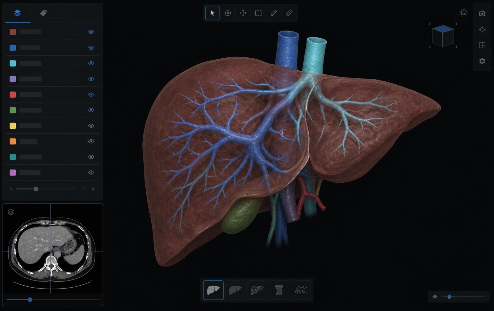
- Interactive 3D model
- Structure visibility controls
- Segmentation colors
- Rotation and zoom
- Vessel visualization
- Lesion visualization
Sophisticated AI underneath. Simple clinical experience on top.
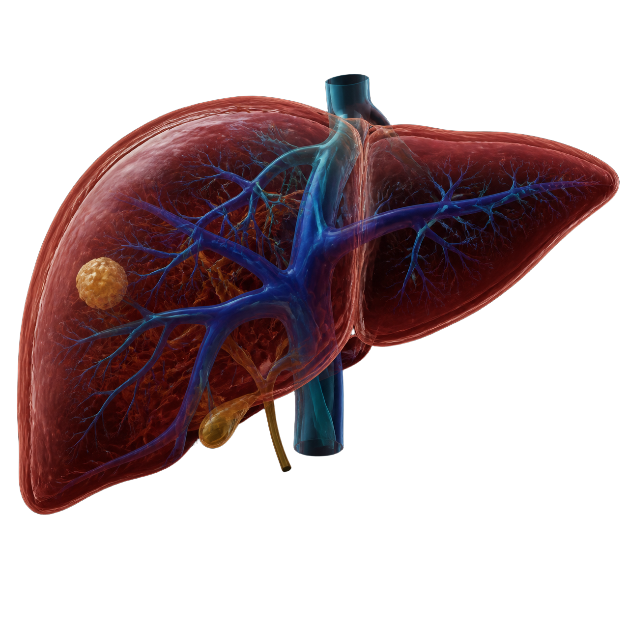
Liver module
Liver planning, reconstructed in 3D.
Patient-specific reconstruction of hepatic anatomy for pre-operative planning, generated from the patient's own CT or MRI examination.
- Liver parenchyma
- Portal venous system
- Hepatic venous system
- Lesions
- Anatomical segmentation
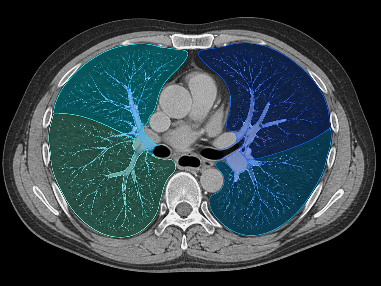
Lung module
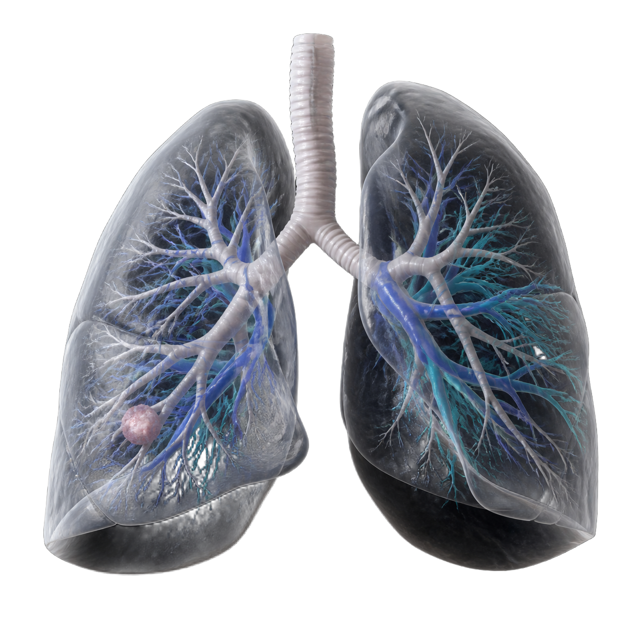
See bronchovascular anatomy in 3D.
Reconstruction of pulmonary anatomy and its spatial relationships, supporting the preparation of thoracic surgical strategy.
- Lungs and lobes
- Bronchi
- Pulmonary arteries
- Pulmonary veins
- Lesions
- Anatomical relationships
Demonstrations
See SPview in action.
Credibility
Built by clinicians.
Designed for surgery.
SPview brings together expertise across radiology, surgery, medical imaging, artificial intelligence and 3D visualization.
- Radiology
- Surgery
- Medical imaging
- Artificial intelligence
- 3D visualization
Our partners
Trusted by surgical teams.
Built with clinicians.
“SPview's 3D liver reconstruction services have been a game-changer for our surgical team. Their technology provides unparalleled clarity and simplicity, helping us identify critical anatomy and plan liver resections with precision. Thanks to SPview, we approach procedures with confidence, knowing we have the most accurate understanding of tumor-vessel proximity and surgical strategy, making them an invaluable partner in our quest for better patient outcomes.”
Prof. Dr. Nouredin MESSAOUDI
HBP Professors, Heelkunde — Brussels, Belgium

“As a young hepatobiliopancreatic surgeon, SPview allows me to achieve better surgical planning, which is essential for the smooth execution of any procedure. It serves as a valuable aid in developing a three-dimensional perspective—an aspect that is particularly challenging in solid organ surgery, where lesions are not always visible beneath the capsule. This tool enables a more confident approach to performing extreme hepatectomies.”
Dr. Lancelot Marique
HBP Surgeon — Adult Liver Transplantation Department, UCL — Brussels, Belgium
“In the modern era, 3D reconstruction has emerged as an invaluable tool, especially with the need for personalized medicine — it can permit surgery and interventions with grace and precision. Concerning HPB surgery, resection planning for the liver presents distinct challenges due to its complex and individually diverse anatomy. This necessitates surgeons to possess a considerable amount of mental visualization ability to adequately plan interventions, a skill that requires years of training and proves challenging to impart. In addition, the surgeon should preserve the functional remnant liver volume, and he has to respect the oncological radicality. At the moment, 3D reconstruction is the prelude of augmented reality — a monumental aid for surgeons, a pivotal ally that bestows upon them invaluable assistance in navigating the complex terrain of liver anatomy.”
Dr. Alessandro Mazzotta
Hepatobiliary Surgeon — Sapienza University of Rome, Italy

Bring 3D surgical planning
into your clinical workflow.
Discover how SPview can transform medical imaging into patient-specific 3D surgical insight directly within your institution.
Dedicated workstation. Local processing. Liver + Lung.